incisive foramen radiograph
The mean width of bone anterior to the incisive canal was 632 143 mm. The purpose of the present study was to review the literature of how to identify the mental foramen mandibular incisive canal and associated neurovascular bundles during.
Maxillary Anterior Landmarks Intraoral Radiographic Anatomy Dentalcare
Inverted Y formation Radiograph comparison Frame 27.

. Light bluewall of maxillary sinus. Mean canal length was 1863 235 mm and males have significantly longer incisive canal than females. Seen as round to ovoid radiolucency located at nearly centre of the mandibular ramus.
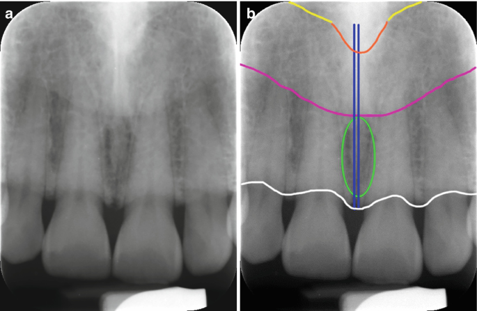
Seen as radiopaque structure on the panoramic radiograph with the shadows superimposed over it. These structures will be completely intact if it is a foramen and interrupted if a periapical pathologic condition is present. Light green ellipselarge incisive foramen.
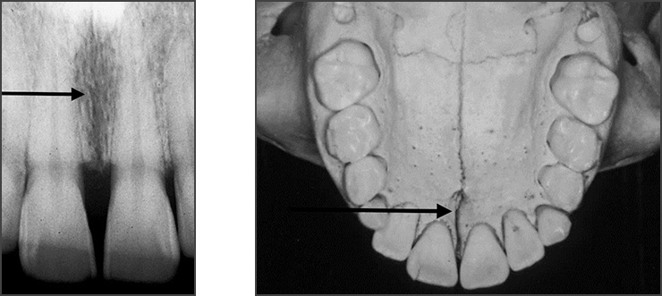
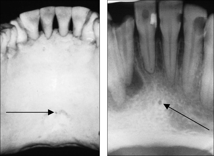
Other normal anatomical landmarks such as the incisive foramen are sometimes present in the area of the midline between the maxillary central incisors and should not be mistaken for an abnormality unless the dimensions are larger than a. Incisive fossa Canine fossa Photograph Frame 28. Familiarity with the incisive foramen is important because.
The small lingual foramen black hole in lower portion of picture as seen on a periapical radiograph of the anterior mandible. However complications may arise due to an extension anterior to the mental foramen that forms the mandible incisive canal MIC. Paired structures are marked on one side only.
URL of Article. The incisive canal located at the midline posterior to the central incisor is an important anatomic structure of this area to be considered while planning for immediate implant placement in maxillary central incisor region. Materials and Methods.
Lateral canals on each side of the midline. Our goal is to evaluate identification of MIC by both panoramic radiograph PAN and cone-beam computed tomography CBCT. Our goal is to evaluate identification of MIC by both panoramic radiograph PAN and cone-beam computed tomography CBCT.
Exit through Foramina of Stenson. Conversely using seven human embryos at weeks seven to 24 Radlanski et al. The purpose of the present study is to assess incisive canal characteristics using CBCT sections.
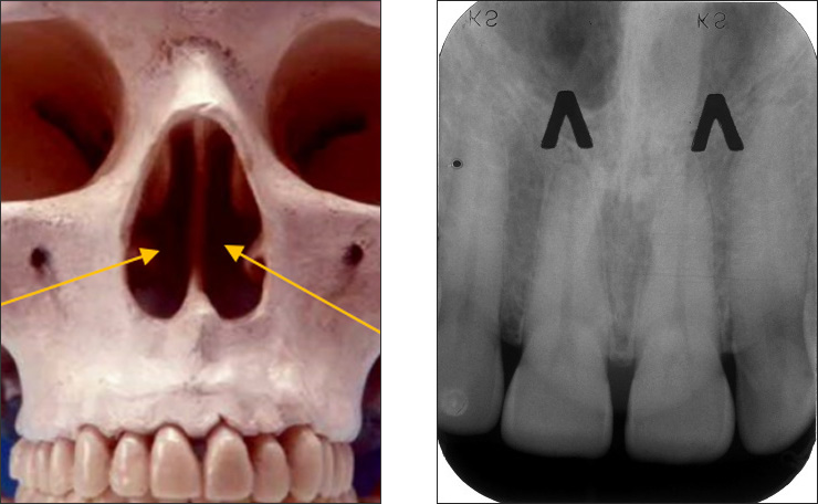
The region between mental foramens is considered as a zone of choice for implants. Purple ellipseinferior nasal concha. Incisive foramen is the opening of the incisive canal located immediately behind the maxillary central incisors.
Tap card to see definition. A b Maxillary occlusal radiograph. Dark bluemidline palatal suture.
The median suture of the palate see figure 3-23 may appear as a radiolucent line extending posteriorly from the alveolar border. It is located in the maxilla in the incisive fossa midline in the palate posterior to the central incisors at the junction of the medial palatine and incisive sutures. Lingula is a tongue shaped radiopaque projection located anterior to the mandibular foramen.
The region between mental foramens is considered as a zone of choice for implants. Another option is to trace the lamina dura and periodontal ligament space. Incisive foramen Median palatine suture Pterygoid plates Pterodactyl gr.
This chapter presents the major landmarks commonly found on conventional dental x-ray images. The lingual foramen gives passage to a single small artery formed. Another radiograph at a different horizontal angu-lation or by testing pulp vitality.
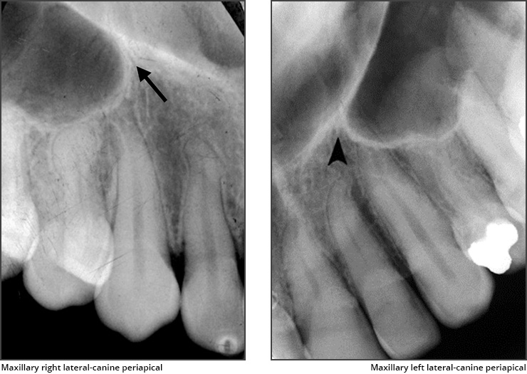
Incisive fossa Canine fossa Radiograph comparison. In radiographs exposed from the region of the cuspid or lateral incisor the incisive foramen may appear as a radiolucency at the apex of one of the incisors. However complications may arise due to an extension anterior to the mental foramen that forms the mandible incisive canal MIC.
The mandibular incisive canal mental foramen and associated neurovascular bundles exist in different locations and possess many variations. The lingual foramen is a small midline opening on the posterior aspect of the symphysis of the mandible just above the mental spine. Panoramic radiograph showing extension of the mental nerve beyond the mental foramen boundary as an intraosseous anterior loop arrows.
Foramen spinosum skull human sphenoid bone ovale boned bones circular immediately pointing probe posterior hole interior through down very which. It can be single or multiple. What is the nasopalatine incisive foramen Click card to see definition.
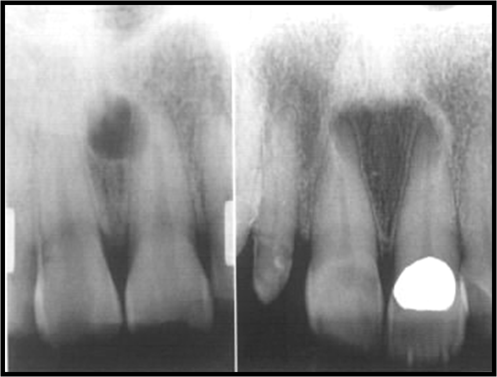
In this case radiographic evaluation and diagnosis help eliminate the need for surgical biopsy andor further imaging studies. Yellowfloor of the nasal cavity. The mean width of the foramen labiopalatally and mesiodistally was 312 094 mm and 323 098 mm respectively.
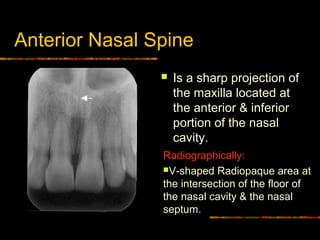
Demonstrated that during embryogenesis the incisive canal was derived from the primary palate within the. On periapical x-ray images the incisive foramen is located in the midline between the roots of the central incisors. 7 Landmarks in the Maxilla Anterior nasal spine Zygomatic process Pterygoid plates Coronoid process of the mandible Nasolabial fold Coronoid Process From the Greek word for Crows Beak.
The incisive foramen also known as nasopalatine foramen or anterior palatine foramen is the oral opening of the nasopalatine canal. Transmit nasopalatine nerves and branches of the descending palatine artery. White ellipseair in nasal cavity.
Furthermore modifications of the cleft palate classifications also include the involvement of the incisive foramen as an extensive form of the submucosal cleft palates 8-11.

Intraoral Radiographs Dr G S Toothpix
Maxillary Anterior Landmarks Intraoral Radiographic Anatomy Dentalcare

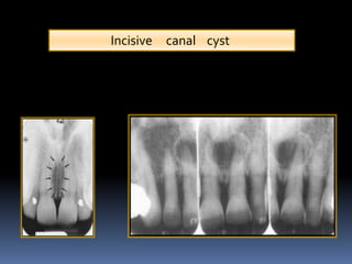
Mouth Incisive Canal Cyst Professional Radiology Outcomes
Incisive Foramen Dr G S Toothpix
Mandibular Anterior Landmarks Intraoral Radiographic Anatomy Dentalcare

Maxillary Anterior Landmarks Intraoral Radiographic Anatomy Continuing Education Course Dentalcare Com

Incisive Canal Radiology Reference Article Radiopaedia Org
Normal Radiographic Anatomical Landmarks

Periapical Radiograph 1 Year After Treatment Bone And Teeth Showing Download Scientific Diagram

Normal Radiographic Anatomical Landmarks
Normal Anatomical Landmarks In Dental X Rays And Cbct Springerlink

File Nasolabial Duct Cyst Jpg Wikipedia
Maxillary Anterior Landmarks Intraoral Radiographic Anatomy Dentalcare

6 Essentials Of Dental Radiographic Analysis And Interpretation Pocket Dentistry

Maxillary Anterior Landmarks Intraoral Radiographic Anatomy Continuing Education Course Dentalcare Com

Measurement Of Nasopalatine Canal Length A Incisive Foramen Diameter Download Scientific Diagram
Radiographic Appearance Of Cysts Part 3 And Scintigraphy Intelligent Dental
Intra Oral Radiographic Anatomical Landmarks

Opg Showing Incisive Foramen And Mental Foramen Download Scientific Diagram


Comments
Post a Comment